Observing the minute world of sperm through a microscope offers a fascinating glimpse into one of the most fundamental processes of life. When viewed under magnification, sperm cells reveal intricate structures and dynamic movements that are invisible to the naked eye. This journey into the microscopic realm not only enhances our understanding of human reproduction but also provides insights into various biological processes. By examining sperm through the lens of a microscope, we unlock a plethora of information that has significant implications for science and medicine.
For many, the idea of viewing sperm under a microscope might seem like an endeavor reserved for scientists in labs. However, with advancements in technology and accessibility, this experience is something that can now be shared by students, educators, and enthusiasts alike. The observation of sperm cells can serve as an excellent educational tool, fostering curiosity and encouraging deeper inquiry into cellular biology. The process of observing sperm through a microscope is not only educational but also an engaging way to witness the marvels of life at its most basic level.
In this article, we will delve into the world of sperm microscopy, exploring its significance in scientific research and education. We will cover the history and evolution of microscopes, the anatomy of sperm, and the various applications of sperm analysis. Through this comprehensive exploration, we aim to shed light on the importance of understanding sperm through a microscope and how this knowledge can contribute to broader scientific and medical advancements.
Table of Contents
- The History and Evolution of Microscopy
- The Anatomy of Sperm Cells
- Types of Microscopes Used in Sperm Observation
- Methods of Sperm Collection and Preparation for Microscopy
- Advanced Microscopic Techniques for Sperm Analysis
- Assessing Sperm Motility and Function
- Understanding Sperm Morphology
- Applications of Sperm Microscopy in Medicine
- The Role of Sperm Microscopy in Fertility Research
- Using Sperm Microscopy as an Educational Tool
- Maintenance and Care of Microscopes
- Safety Considerations in Sperm Microscopy
- The Future of Sperm Microscopy and Technological Innovations
- Frequently Asked Questions
- Conclusion
The History and Evolution of Microscopy
The history of microscopy dates back to the late 16th century when the first compound microscopes were invented. These early instruments were rudimentary, consisting of a series of lenses that magnified small objects. The invention of the microscope is attributed to Zacharias Janssen, a Dutch spectacle maker, who, along with his father Hans, developed the first compound microscope around 1590. This innovation paved the way for future developments in the field of microscopy.
As technology advanced, so did the design and capabilities of microscopes. In the 17th century, Antonie van Leeuwenhoek, a Dutch scientist, made significant contributions to microscopy by improving lens quality and magnification. Van Leeuwenhoek's microscopes were simple but powerful, allowing him to observe microorganisms, including sperm cells, for the first time. His observations laid the groundwork for the study of microbiology and cellular biology.
The 19th century saw further advancements with the introduction of achromatic lenses, which corrected color distortions and improved image clarity. This period also marked the development of the first electron microscopes, which utilized electron beams instead of light to achieve higher magnifications and resolution. These technological advancements have allowed scientists to observe sperm cells in unprecedented detail, providing valuable insights into their structure and function.
Today, microscopy continues to evolve with the advent of digital imaging, fluorescence microscopy, and confocal microscopy. These state-of-the-art techniques enable researchers to explore the intricate world of sperm cells and other microscopic entities with remarkable precision. As our understanding of microscopy advances, so too does our ability to unlock the secrets of life at the cellular level.
The Anatomy of Sperm Cells
Sperm cells, or spermatozoa, are the male reproductive cells responsible for fertilizing the female egg. They are highly specialized cells, designed to deliver the male's genetic material to the egg. Understanding the anatomy of sperm cells is crucial for studying their function and role in reproduction.
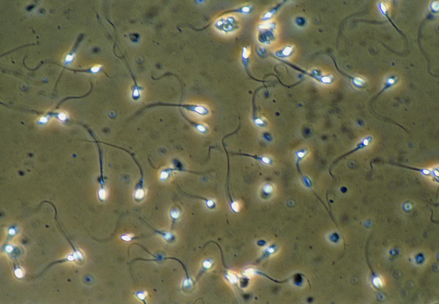
Sperm cells consist of three main parts: the head, midpiece, and tail. The head contains the nucleus, which houses the genetic material, and is covered by an acrosome, a cap-like structure that contains enzymes necessary for penetrating the egg. The acrosome plays a vital role in fertilization by facilitating the sperm's entry into the egg.
The midpiece of the sperm is packed with mitochondria, the energy-producing organelles of the cell. These mitochondria provide the energy required for the sperm's motility, enabling it to swim through the female reproductive tract to reach the egg. The midpiece is a crucial component for the sperm's movement and overall function.
The tail, or flagellum, is a whip-like structure that propels the sperm forward. It is composed of microtubules arranged in a specific pattern, allowing for efficient movement. The tail's ability to generate movement is essential for successful fertilization, as it enables the sperm to navigate through the challenging environment of the female reproductive system.
Understanding the anatomy of sperm cells provides a foundation for studying their behavior and function. By observing sperm under a microscope, researchers can gain insights into their motility, morphology, and overall health, which are critical factors in reproductive biology.
Types of Microscopes Used in Sperm Observation
The observation of sperm cells requires specialized microscopes that offer high magnification and resolution. Several types of microscopes are commonly used in sperm analysis, each with unique features and capabilities.
Light Microscopes
Light microscopes, also known as optical microscopes, are the most commonly used instruments for observing sperm cells. They utilize visible light to illuminate the specimen and magnify the image through a series of lenses. There are different types of light microscopes, including compound microscopes and phase-contrast microscopes.
Compound microscopes are widely used in educational settings and laboratories for basic sperm observation. They provide sufficient magnification to view the overall structure and movement of sperm cells. Phase-contrast microscopes enhance the contrast of transparent specimens, making them ideal for observing live sperm cells and assessing their motility.
Electron Microscopes
Electron microscopes offer much higher magnification and resolution compared to light microscopes. They use electron beams to create detailed images of sperm cells, revealing intricate structures that are not visible with light microscopy. There are two main types of electron microscopes: transmission electron microscopes (TEM) and scanning electron microscopes (SEM).
TEMs are used to observe the internal structures of sperm cells, providing detailed images of the nucleus, acrosome, and other cellular components. SEMs, on the other hand, produce three-dimensional images of the sperm's surface, allowing researchers to study its morphology and surface features.
Fluorescence Microscopes
Fluorescence microscopy is a powerful technique that uses fluorescent dyes to label specific components of sperm cells. These dyes emit light when exposed to certain wavelengths, allowing researchers to visualize specific structures and processes within the cells. Fluorescence microscopes are particularly useful for studying sperm motility, acrosome reaction, and other dynamic processes.
Each type of microscope offers unique advantages and is chosen based on the specific requirements of the study. By employing a combination of these techniques, researchers can gain a comprehensive understanding of sperm cells and their functions.
Methods of Sperm Collection and Preparation for Microscopy
Collecting and preparing sperm samples for microscopy is a critical step in sperm analysis. The methods used for collection and preparation can significantly impact the quality and reliability of the observations. There are several techniques for collecting sperm samples, each suited to different research and clinical purposes.
Semen Collection
The most common method of obtaining sperm samples is through semen collection. This procedure involves collecting ejaculate through masturbation or coitus interruptus, where the male withdraws before ejaculation. In clinical settings, semen collection is typically done in a private room, ensuring the sample is fresh and uncontaminated.
Testicular Biopsy
In cases where semen is not available, such as in individuals with azoospermia (absence of sperm in ejaculate), a testicular biopsy may be performed. This procedure involves obtaining a small tissue sample from the testes, which is then processed to extract sperm cells. Testicular biopsy is a more invasive method but can provide valuable information in specific cases.
Sperm Preparation for Microscopy
Once the sperm sample is collected, it must be prepared for microscopy to ensure accurate observations. The preparation process typically involves dilution, washing, and staining of the sample.
Dilution: The sperm sample is diluted with a suitable medium to reduce the concentration of sperm cells, allowing for better visibility under the microscope.
Washing: The sample is washed to remove debris, seminal plasma, and other contaminants that may interfere with the observation.
Staining: Staining is an optional step that involves adding a dye to the sample to enhance the contrast of the sperm cells. Common stains used in sperm microscopy include eosin-nigrosin and Papanicolaou stain.
Proper collection and preparation of sperm samples are essential for accurate and meaningful observations. By following standardized protocols, researchers can ensure the reliability and reproducibility of their findings.
Advanced Microscopic Techniques for Sperm Analysis
In recent years, advancements in microscopic techniques have revolutionized the field of sperm analysis. These cutting-edge methods provide unprecedented insights into sperm behavior, structure, and function, enhancing our understanding of reproductive biology.
Computer-Assisted Sperm Analysis (CASA)
Computer-Assisted Sperm Analysis (CASA) is a sophisticated technique that uses computer software to analyze sperm motility and concentration. CASA systems capture video footage of sperm samples and utilize algorithms to track and measure the movement patterns of individual sperm cells. This technology provides precise and objective data on sperm motility, which is crucial for assessing male fertility.
Confocal Microscopy
Confocal microscopy is a powerful imaging technique that uses laser light to scan and create high-resolution, three-dimensional images of sperm cells. This method allows researchers to visualize the internal structures of sperm cells with exceptional clarity, providing insights into their morphology and function. Confocal microscopy is particularly useful for studying sperm morphology and identifying structural abnormalities.
Fluorescence Lifetime Imaging Microscopy (FLIM)
Fluorescence Lifetime Imaging Microscopy (FLIM) is an advanced technique that measures the time it takes for fluorescent molecules to emit light after being excited by a laser. This method provides information about the biochemical environment within sperm cells, allowing researchers to study cellular processes such as energy metabolism and signaling pathways. FLIM is a valuable tool for investigating sperm function and viability.
Atomic Force Microscopy (AFM)
Atomic Force Microscopy (AFM) is a cutting-edge technique that uses a sharp probe to scan the surface of sperm cells at the nanometer scale. AFM provides detailed images of the sperm's surface topography, enabling researchers to study its morphology and mechanical properties. This technique is particularly useful for examining the structure of the sperm head and tail.
These advanced microscopic techniques offer new possibilities for sperm analysis, providing valuable insights into the complex biology of sperm cells. By leveraging these technologies, researchers can enhance our understanding of male fertility and reproductive health.
Assessing Sperm Motility and Function
Sperm motility, or the ability of sperm cells to move effectively, is a critical factor in male fertility. Assessing sperm motility is essential for evaluating the potential of sperm to reach and fertilize the egg. There are several methods for assessing sperm motility and function, each providing valuable information about the health and viability of sperm cells.
Visual Assessment
The simplest method of assessing sperm motility is through visual observation under a light microscope. In this approach, a semen sample is placed on a microscope slide, and the movement patterns of sperm cells are observed. Sperm motility is typically classified into categories such as progressive motility (forward movement), non-progressive motility (random movement), and immotility (no movement).
Computer-Assisted Sperm Analysis (CASA)
As mentioned earlier, Computer-Assisted Sperm Analysis (CASA) is a sophisticated technique that provides objective and quantitative data on sperm motility. CASA systems capture video footage of sperm samples and use algorithms to analyze the movement patterns of individual sperm cells. This technology offers precise measurements of parameters such as velocity, linearity, and amplitude of lateral head displacement.
Sperm Function Tests
In addition to motility assessments, sperm function tests are used to evaluate the ability of sperm cells to perform specific functions necessary for fertilization. These tests may include:
- Acrosome Reaction Test: This test assesses the ability of sperm cells to undergo the acrosome reaction, a crucial step in fertilization where enzymes are released to penetrate the egg's protective layers.
- Zona Binding Test: This test evaluates the ability of sperm cells to bind to the zona pellucida, the outer layer of the egg, which is essential for successful fertilization.
- Hypo-Osmotic Swelling Test: This test assesses the integrity of the sperm cell membrane, which is important for maintaining cell viability and function.
Assessing sperm motility and function is a critical component of male fertility evaluation. By combining visual observation, CASA, and sperm function tests, researchers and clinicians can gain a comprehensive understanding of sperm health and potential reproductive outcomes.
Understanding Sperm Morphology
Sperm morphology, or the shape and structure of sperm cells, is an essential aspect of male fertility assessment. Abnormal sperm morphology can significantly impact the ability of sperm to reach and fertilize the egg. Understanding sperm morphology involves examining the size, shape, and structure of sperm cells to identify any abnormalities that may affect reproductive outcomes.
Normal Sperm Morphology
Normal sperm cells have a characteristic shape, consisting of an oval head, a midpiece, and a long tail. The head of the sperm is typically smooth and symmetrical, with a well-defined acrosome and nucleus. The midpiece is cylindrical and contains mitochondria, while the tail is slender and whip-like, facilitating movement.
Abnormal Sperm Morphology
Abnormal sperm morphology refers to any deviations from the typical shape and structure of sperm cells. Common abnormalities include:
- Head Abnormalities: These may include large or small heads, tapered heads, or heads with irregular shapes.
- Midpiece Abnormalities: These may include swollen or irregular midpieces, which can affect sperm motility.
- Tail Abnormalities: These may include coiled, bent, or multiple tails, which can impair movement.
Abnormal sperm morphology can result from a variety of factors, including genetic defects, environmental exposures, lifestyle factors, and underlying medical conditions. Identifying and understanding these abnormalities is crucial for diagnosing male infertility and developing appropriate treatment strategies.
Assessment of Sperm Morphology
Sperm morphology is typically assessed through microscopic examination of stained semen samples. The World Health Organization (WHO) provides guidelines for the assessment of sperm morphology, including criteria for categorizing sperm cells as normal or abnormal. The percentage of normal sperm cells in a sample is an important parameter in fertility evaluations.
Understanding sperm morphology is essential for evaluating male fertility and identifying potential causes of infertility. By examining the shape and structure of sperm cells, researchers and clinicians can gain valuable insights into reproductive health and develop targeted interventions to improve fertility outcomes.
Applications of Sperm Microscopy in Medicine
Sperm microscopy has a wide range of applications in medicine, providing valuable insights into male fertility, reproductive health, and broader medical conditions. By observing sperm cells under a microscope, researchers and clinicians can gain critical information that informs diagnosis, treatment, and research.
Diagnosis of Male Infertility
Sperm microscopy is a fundamental tool in the diagnosis of male infertility. By assessing parameters such as sperm count, motility, and morphology, clinicians can identify potential causes of infertility and develop appropriate treatment strategies. Sperm analysis is often the first step in fertility evaluations and can help guide subsequent interventions.
Assisted Reproductive Technologies (ART)
In the context of assisted reproductive technologies (ART), sperm microscopy plays a critical role in the selection of sperm cells for procedures such as in vitro fertilization (IVF) and intracytoplasmic sperm injection (ICSI). By identifying the healthiest and most motile sperm cells, clinicians can improve the chances of successful fertilization and pregnancy outcomes.
Research on Reproductive Health
Sperm microscopy is an invaluable tool for research on reproductive health and male fertility. By studying sperm cells, researchers can gain insights into the underlying mechanisms of fertility, the impact of environmental and lifestyle factors, and the development of new diagnostic and therapeutic approaches. Sperm microscopy contributes to a deeper understanding of reproductive biology and supports the advancement of fertility treatments.
Investigation of Medical Conditions
Beyond fertility assessments, sperm microscopy can be used to investigate medical conditions that affect sperm production and function. Conditions such as varicocele, hormonal imbalances, and genetic disorders can impact sperm health, and microscopic analysis can aid in their diagnosis and management. Sperm microscopy provides valuable information for understanding the impact of these conditions on reproductive health.
The applications of sperm microscopy in medicine are vast and varied, offering critical insights into male fertility and reproductive health. By leveraging the power of microscopy, researchers and clinicians can enhance our understanding of reproductive biology and improve outcomes for individuals facing fertility challenges.
The Role of Sperm Microscopy in Fertility Research
Sperm microscopy is a cornerstone of fertility research, providing essential insights into the biology of sperm cells and their role in reproduction. By observing sperm under a microscope, researchers can investigate the factors that influence fertility and develop new strategies for improving reproductive outcomes.
Sperm Motility and Fertility
Sperm motility is a critical factor in male fertility, and sperm microscopy allows researchers to study the movement patterns of sperm cells in detail. By assessing parameters such as velocity, linearity, and amplitude of lateral head displacement, researchers can gain insights into the factors that affect sperm motility and identify potential interventions to improve it. Sperm microscopy is instrumental in understanding the relationship between motility and fertility.
Sperm Morphology and Fertility
Sperm morphology is another key factor in fertility, and sperm microscopy is essential for assessing the shape and structure of sperm cells. By identifying abnormalities in sperm morphology, researchers can investigate the underlying causes and develop targeted interventions to improve fertility outcomes. Sperm microscopy is a critical tool for understanding the impact of morphology on reproductive success.
Impact of Environmental and Lifestyle Factors
Sperm microscopy is also used to study the impact of environmental and lifestyle factors on male fertility. By observing changes in sperm parameters such as motility, morphology, and concentration, researchers can investigate the effects of factors such as diet, exercise, pollution, and stress on reproductive health. Sperm microscopy provides valuable insights into the relationship between lifestyle and fertility.
Development of New Fertility Treatments
Sperm microscopy plays a crucial role in the development of new fertility treatments and interventions. By studying the biology of sperm cells, researchers can identify potential targets for therapeutic interventions and develop new diagnostic tools and treatments for male infertility. Sperm microscopy supports the advancement of fertility research by providing critical insights into the mechanisms of reproduction.
The role of sperm microscopy in fertility research is invaluable, offering essential insights into the factors that influence male fertility and reproductive health. By leveraging the power of microscopy, researchers can enhance our understanding of reproductive biology and develop new strategies for improving fertility outcomes.
Using Sperm Microscopy as an Educational Tool
Sperm microscopy is not only a valuable tool for research and clinical applications but also an excellent educational tool for teaching cellular biology and reproductive health. By observing sperm cells under a microscope, students and educators can gain a deeper understanding of the biology of reproduction and the factors that influence fertility.
Hands-On Learning
Sperm microscopy provides a hands-on learning experience that engages students and fosters curiosity. By observing sperm cells in real-time, students can witness the dynamic behavior and intricate structures of these cells, enhancing their understanding of cellular biology and reproduction. Sperm microscopy offers a unique opportunity for experiential learning and inquiry-based education.
Understanding Reproductive Health
Sperm microscopy can also be used to educate students about reproductive health and the factors that influence fertility. By studying sperm cells, students can learn about the biology of reproduction, the impact of lifestyle and environmental factors on fertility, and the importance of healthy reproductive practices. Sperm microscopy supports comprehensive education on reproductive health and wellness.
Encouraging Scientific Inquiry
Sperm microscopy encourages scientific inquiry and critical thinking by challenging students to observe, analyze, and interpret their findings. By investigating the behavior and structure of sperm cells, students can develop essential skills in observation, data analysis, and scientific reasoning. Sperm microscopy fosters a culture of curiosity and inquiry in the classroom.
Using sperm microscopy as an educational tool offers valuable opportunities for teaching cellular biology and reproductive health. By engaging students in hands-on learning and scientific inquiry, educators can enhance their understanding of the biology of reproduction and the factors that influence fertility.
Maintenance and Care of Microscopes
Proper maintenance and care of microscopes are essential for ensuring their longevity and optimal performance. By following recommended maintenance practices, users can keep their microscopes in excellent condition and prevent damage or deterioration.
Cleaning and Storage
Regular cleaning is crucial for maintaining the performance of microscopes. Dust and debris can accumulate on lenses and other components, affecting image quality. Users should clean lenses with lens paper or a microfiber cloth and use compressed air to remove dust. Microscopes should be stored in a dry, dust-free environment, covered with a protective cover when not in use.
Handling and Usage
Proper handling and usage of microscopes are critical for preventing damage. Users should hold the microscope by the arm and base, avoid touching lenses with fingers, and use appropriate lighting settings to prevent overheating. Microscopes should be operated according to the manufacturer's instructions to ensure safe and effective use.
Routine Maintenance
Routine maintenance includes checking and adjusting mechanical components, such as the stage and focus knobs, to ensure smooth operation. Users should also inspect electrical components, such as the light source and power supply, for any signs of wear or damage. Regular calibration and alignment are recommended to maintain optimal performance.
By following recommended maintenance and care practices, users can ensure the longevity and performance of their microscopes. Proper maintenance supports accurate and reliable observations, enhancing the quality of research and education.
Safety Considerations in Sperm Microscopy
Safety is a critical consideration in sperm microscopy, ensuring the well-being of researchers, students, and the environment. By following recommended safety practices, users can prevent accidents and protect themselves from potential hazards.
Laboratory Safety
Laboratory safety protocols should be followed to prevent accidents and injuries. Users should wear appropriate personal protective equipment (PPE), such as lab coats, gloves, and safety goggles, and follow safe handling and disposal procedures for biological samples and chemicals. Laboratories should be equipped with safety equipment, such as eyewash stations and fire extinguishers, and users should be trained in emergency procedures.
Sample Handling
Proper handling of sperm samples is essential for ensuring safety and preventing contamination. Samples should be collected and processed in a clean, controlled environment, and users should follow aseptic techniques to prevent contamination. Proper labeling and storage of samples are also important for maintaining safety and accuracy.
Chemical Safety
Chemicals used in sperm microscopy, such as stains and fixatives, can pose potential hazards. Users should follow safety guidelines for handling and disposing of chemicals, including using appropriate PPE and working in a well-ventilated area. Material Safety Data Sheets (MSDS) should be available for all chemicals used in the laboratory.
By following recommended safety practices, users can ensure a safe and productive environment for sperm microscopy. Safety is a critical consideration in research and education, protecting the well-being of individuals and the environment.
The Future of Sperm Microscopy and Technological Innovations
The field of sperm microscopy continues to evolve, driven by technological advancements and innovations that enhance our understanding of reproductive biology. As new techniques and technologies emerge, the future of sperm microscopy holds exciting possibilities for research, diagnostics, and education.
Advancements in Imaging Technology
Advancements in imaging technology, such as high-resolution digital imaging and three-dimensional microscopy, are transforming the field of sperm microscopy. These technologies offer unprecedented insights into the structure and function of sperm cells, enhancing our understanding of their biology and role in reproduction. The future of sperm microscopy will be shaped by continued innovations in imaging technology.
Integration of Artificial Intelligence
The integration of artificial intelligence (AI) in sperm microscopy is opening new avenues for research and diagnostics. AI algorithms can analyze large datasets, identify patterns, and provide insights into sperm behavior and function. AI-powered sperm analysis systems offer the potential for more accurate and efficient assessments, supporting advancements in fertility research and diagnostics.
Development of New Diagnostic Tools
The future of sperm microscopy will be marked by the development of new diagnostic tools and techniques for assessing male fertility. These innovations will provide more comprehensive and accurate assessments, improving the diagnosis and treatment of male infertility. The development of new diagnostic tools will be driven by advancements in technology and a deeper understanding of reproductive biology.
The future of sperm microscopy holds exciting possibilities for enhancing our understanding of reproductive biology and improving fertility outcomes. As new technologies and innovations emerge, the field of sperm microscopy will continue to evolve, offering new insights and opportunities for research, diagnostics, and education.
Frequently Asked Questions
1. What is the purpose of observing sperm through a microscope?
Observing sperm through a microscope allows researchers and clinicians to assess sperm motility, morphology, and concentration, which are critical factors in evaluating male fertility. Microscopy provides valuable insights into the health and function of sperm cells, supporting the diagnosis and treatment of male infertility.
2. What types of microscopes are used for sperm analysis?
Several types of microscopes are used for sperm analysis, including light microscopes, electron microscopes, and fluorescence microscopes. Each type offers unique advantages, with light microscopes commonly used for basic observations, electron microscopes providing high-resolution images, and fluorescence microscopes allowing for specific labeling of cellular components.
3. How is sperm motility assessed under a microscope?
Sperm motility is typically assessed through visual observation under a light microscope or using computer-assisted sperm analysis (CASA) systems. These methods evaluate the movement patterns of sperm cells, providing information on their velocity, linearity, and overall motility, which are important for assessing male fertility.
4. What factors can affect sperm morphology?
Sperm morphology can be affected by a variety of factors, including genetic defects, environmental exposures, lifestyle factors, and underlying medical conditions. Abnormalities in sperm morphology can impact fertility, making it essential to identify and understand these factors when evaluating male reproductive health.
5. How can sperm microscopy be used as an educational tool?
Sperm microscopy can be used as an educational tool to teach cellular biology and reproductive health. By observing sperm cells under a microscope, students can gain a deeper understanding of the biology of reproduction, the factors that influence fertility, and the importance of healthy reproductive practices.
6. What are the future prospects for sperm microscopy?
The future of sperm microscopy is promising, with advancements in imaging technology, artificial intelligence, and new diagnostic tools enhancing our understanding of reproductive biology. These innovations offer exciting possibilities for research, diagnostics, and education, improving fertility outcomes and advancing the field of reproductive health.
Conclusion
Sperm microscopy offers a remarkable window into the microscopic world of reproduction, providing invaluable insights into the biology and function of sperm cells. From assessing motility and morphology to advancing fertility research and education, the observation of sperm through a microscope plays a critical role in understanding male fertility and reproductive health.
As technology continues to evolve, the field of sperm microscopy will undoubtedly expand, offering new possibilities for research, diagnosis, and treatment. By leveraging the power of microscopy, researchers, clinicians, and educators can enhance our understanding of reproductive biology and improve outcomes for individuals facing fertility challenges.
The journey into the world of sperm microscopy is a testament to the wonders of science and the endless pursuit of knowledge. As we continue to explore the intricate and dynamic world of sperm cells, we are reminded of the beauty and complexity of life at its most fundamental level.